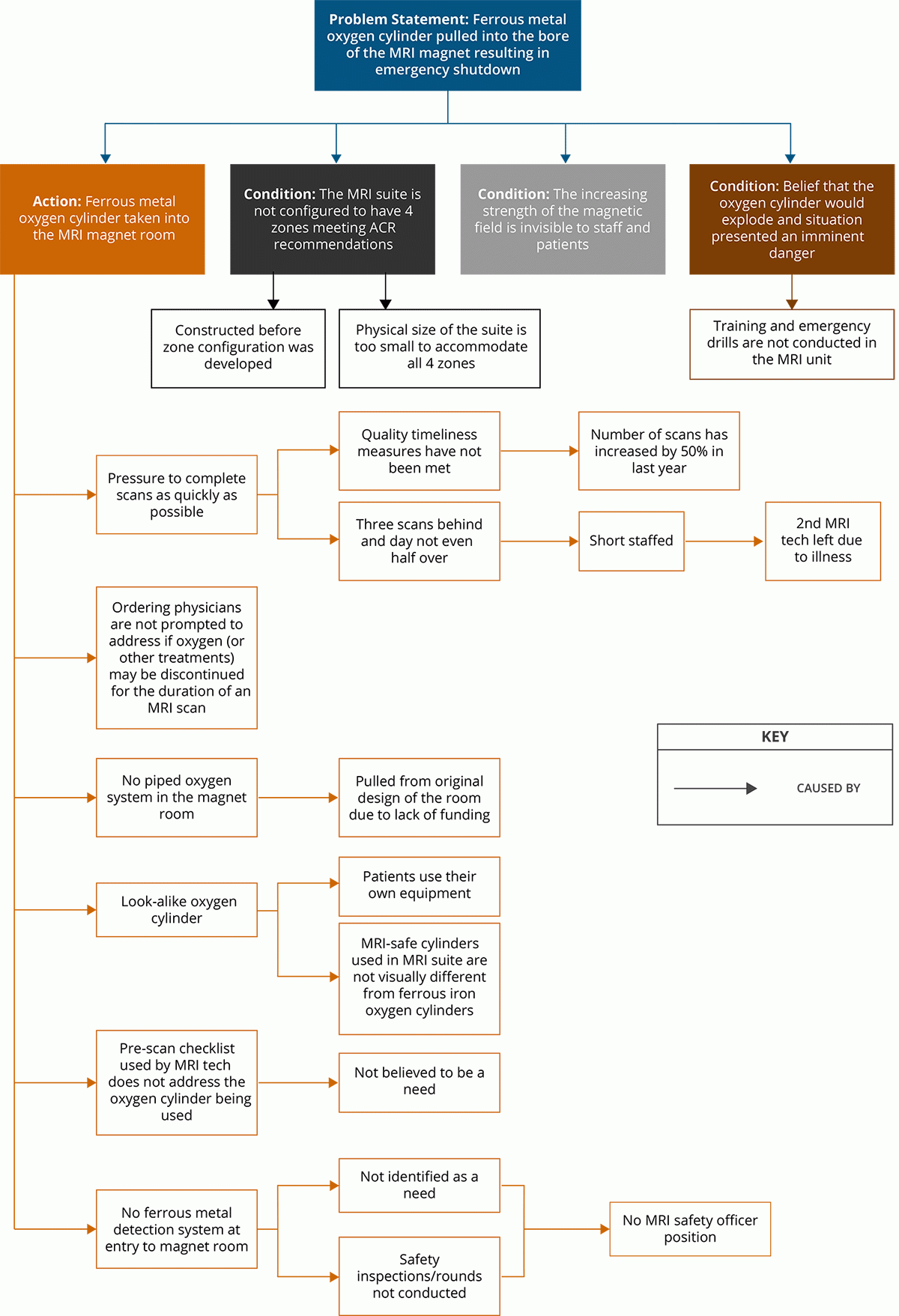
Describe what happened in a series of numbered boxes. Beside each box, add information gleaned from the team’s inquiry that answer questions such as “how” or “why” this step is relevant to the adverse event or near miss under analysis.
1. Patient (JP) has COPD and is on oxygen (2 lpm) and requires knee surgery. | JP could have had his oxygen therapy discontinued for the duration of the MR scan without causing complications. |
2. JP reports for a previously scheduled outpatient MRI. | There were no notes in the EMR about the patient being on oxygen or whether it could be discontinued for the duration of the scan. JP was not given any informational material about the scan. |
3. JP arrives at the MRI suite with his oxygen cylinder. | The oxygen cylinder that JP is using looks identical to the MRI safe oxygen cylinders used in the MRI suite. The receptionist didn’t question the oxygen cylinder as it wasn’t part of the job but sometimes he did to help out; the MRI tech thought that the cylinder had already been switched to an MRI safe cylinder. |
4. JP checks in and is asked to change out of his street clothes and put on scrubs. He was also asked to remove any chains, watches, and jewelry. | It is the policy to change into scrubs. A changing room is available along with lockers for patient use. |
5. The MR tech escorts JP from the changing room to just outside the entrance of the magnet room. JP still has his oxygen cylinder with him. | The MR suite is not designed in accordance with the four zone, dirty (ferrous metal) to clean (no ferrous metal) concept advocated by the American College of Radiology. |
6. The MR tech questions JP about jewelry, implants, patches, etc. | A standardized form/checklist is used to question all patients about metal objects they may be carrying or have implanted; oxygen cylinders are supposed to be provided by the facility and are not on the form. The protocol is for objects such as gurneys, wheelchairs, oxygen cylinders to be switched out to MR safe or MR conditional equipment before the MR tech meets the patient. |
7. The MR tech is called away in the middle of questioning JP and returns a few minutes later to finish. | The tech was called away to answer a question from a physician; while he was taking care of this the clerk reminded him that they were 3 appointments behind and that maybe they could get caught up over lunch. The day before staff had been told that their new quality measure was timeliness and patient waiting times. The MR unit was short staffed on this day due to an illness. |
8. The MR tech asks JP to follow him into the magnet room. JP does so pulling the oxygen cylinder behind him. | A ferrous metal detector is not provided at the entrance into the magnet room and hand held scanners are not used. A sign on the door warns to remove all metal before entering. The magnet room does not have piped in oxygen. |
9. As JP approaches the MR table the oxygen cylinder is drawn into the bore of the magnet narrowly missing the tech as it flies by him. | There are no visual clues or indicators in the room to warn individuals about the increasing magnetic field. |
10. The tech activates the emergency MRI shutdown. Engineering/Facilities are called. | The tech thought that the oxygen cylinder could explode. He was not aware of the possible safety consequences or equipment damage when the magnet is quenched by instituting an emergency MRI shutdown. The tech did not recall any training being done on emergency shutdowns. |
11. A vendor is contacted, the MR unit helium is recharged and the cracked cowling is replaced. | |
12. MRI service is resumed approximately 5 days after the event occurred. |
All events appearing in this table are fictitious. Any resemblance to real events is purely coincidental.
Based on the Cause and Effect Diagramming Model from Apollo Root Cause Analysis: A New Way of Thinking by Dean L. Gano (Apollonian Publications, 1999.)
Then add causation factors to the events as illustrated below.